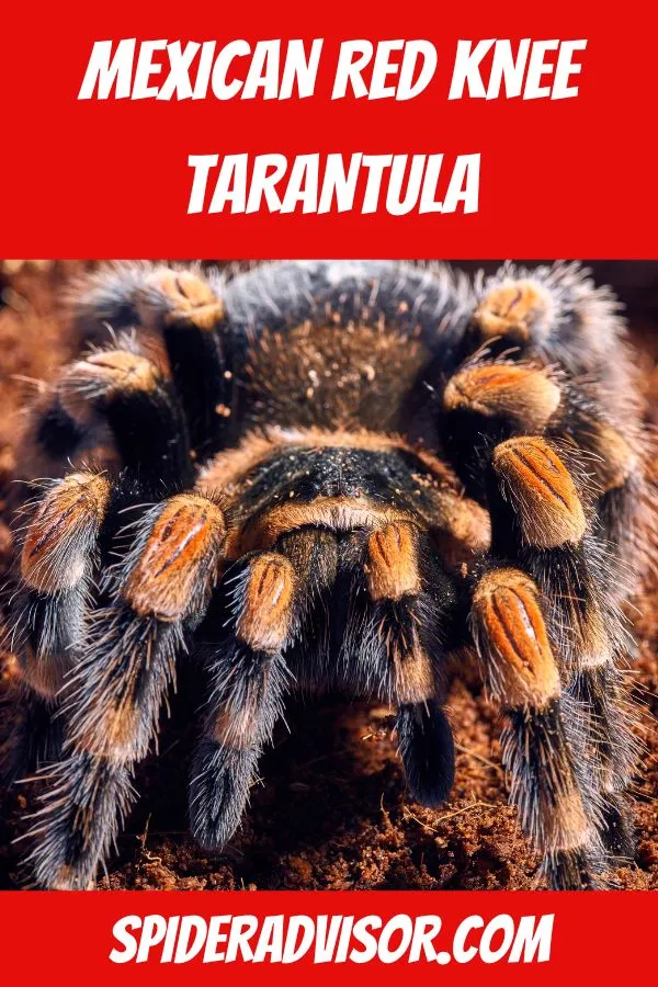
The Mexican Red Knee Tarantula (Brachypelma hamorii) is a captivating creature, beloved by both novice and experienced arachnid enthusiasts. Understanding the intricate body parts of this stunning spider is key to appreciating its unique biology and ensuring its well-being. This guide unveils the top 5 essential facts about the Mexican Red Knee Tarantula’s anatomy, providing valuable insights for anyone fascinated by these remarkable animals. From the fundamental building blocks to the specialized structures that enable survival, get ready to explore the fascinating world of the Mexican Red Knee Tarantula’s body parts.
Cephalothorax The Body’s Core
The cephalothorax, also known as the prosoma, is the fused head and thorax region of the tarantula. This crucial section serves as the central command center, housing vital organs and sensory structures. It’s a robust, well-protected area, crucial for the spider’s survival. The cephalothorax’s exterior is covered by a hardened exoskeleton, providing both protection and structural support. This tough outer shell is essential for shielding the delicate internal organs from harm. The cephalothorax of a Mexican Red Knee Tarantula is typically dark in color, often with contrasting patterns that contribute to its striking appearance and camouflage capabilities. This part of the body is incredibly important for the overall health and functionality of the tarantula.
The Carapace Protection and Structure
The carapace is the dorsal (upper) surface of the cephalothorax, acting as a protective shield. It’s a tough, plate-like structure made of chitin, the same material that forms the exoskeleton. The carapace’s primary function is to protect the spider’s vital organs, including the brain, heart, and other crucial internal components. The shape and texture of the carapace can vary slightly between different tarantula species. In the Mexican Red Knee, the carapace is typically adorned with distinctive markings, adding to the spider’s visual appeal. The carapace is also a site of muscle attachments, which are essential for the spider’s movement and other bodily functions. Its robust structure ensures that the tarantula remains safe from potential threats in its environment, and it’s a critical part of the spider’s anatomy.
Eyesight and Sensory Organs
While tarantulas don’t have the same visual acuity as humans, their eyesight is still essential for survival. They possess eight eyes, arranged in two rows on the front of the cephalothorax. These eyes primarily detect movement, light, and shadow, helping them to navigate and hunt. In addition to eyes, tarantulas have other sensory organs, including slit sensilla, which are tiny slits in the exoskeleton that detect vibrations and changes in air pressure. These help the tarantula sense the presence of prey or potential threats. The combination of sight and other sensory inputs allows the Mexican Red Knee Tarantula to perceive its surroundings, making it an effective predator. This complex sensory system is crucial for survival in its natural habitat, allowing it to react quickly to changes in its environment.
The Chelicerae Fangs and Venom
The chelicerae are the mouthparts located at the front of the cephalothorax, housing the fangs. These fangs are used to inject venom into prey, immobilizing them for feeding. The size of the fangs can vary depending on the tarantula’s size and species. The chelicerae are incredibly strong and are capable of piercing the exoskeletons of insects and other small creatures. The venom itself is a complex mixture of toxins that work to paralyze or kill prey. The Mexican Red Knee Tarantula’s venom is not considered highly dangerous to humans, but a bite can still cause pain and localized swelling. Understanding the function of the chelicerae and fangs is crucial for responsible tarantula ownership, including handling safety and recognizing the potential for defense.
Abdomen Digestive and Reproductive System
The abdomen is the posterior (rear) section of the tarantula, and it’s connected to the cephalothorax by a narrow pedicel. This section is primarily responsible for housing the digestive and reproductive organs. The abdomen is typically larger and more flexible than the cephalothorax, allowing for expansion as the tarantula feeds. The exoskeleton of the abdomen is also covered in hairs, which can play a role in sensory perception and defense. The abdomen’s internal structure is intricate, with the digestive system efficiently processing food and the reproductive organs contributing to the species’ continuation. Understanding the abdomen’s functions is essential for knowing the health and reproductive status of the tarantula.
Book Lungs Respiration and Oxygen Intake
Tarantulas breathe using book lungs, unique respiratory organs located in the abdomen. These structures resemble the pages of a book, providing a large surface area for gas exchange. The book lungs absorb oxygen from the air and transfer it to the tarantula’s circulatory system, while also removing carbon dioxide. The number of book lungs can vary among different tarantula species. The book lungs are protected by the exoskeleton, which has small openings called spiracles that allow air to enter. The efficiency of the book lungs is vital to the tarantula’s ability to move, hunt, and perform all the necessary biological functions. Damage or dysfunction of the book lungs can severely impact the spider’s health, highlighting their significance.
Spinnerets Silk Production
Spinnerets are specialized structures located at the end of the abdomen, used for producing silk. Tarantulas have multiple spinnerets, each capable of producing different types of silk with varied properties. This silk is used for several purposes, including building burrows, creating draglines, and constructing egg sacs. The spinnerets are connected to silk glands within the abdomen, where the silk proteins are synthesized. The spider controls the amount and type of silk it produces, adjusting its output according to the needs of the situation. The ability to produce silk is fundamental to the tarantula’s survival, providing the tools for shelter, movement, and reproduction. The silk is a crucial element in the spider’s ecological niche.
Legs and Pedipalps Movement and Sensory Function
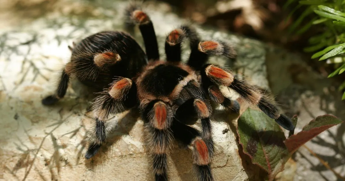
The Mexican Red Knee Tarantula, like all spiders, has eight legs. These legs are not just for walking; they are also used for climbing, hunting, and sensing the environment. The legs are covered in sensory hairs that detect vibrations, air currents, and other stimuli. This detailed sensory information allows the tarantula to perceive its surroundings and react to potential threats or opportunities. The legs are attached to the cephalothorax and are controlled by muscles within the leg segments. In addition to legs, the tarantula has two pedipalps, which are located near the mouth and serve multiple functions, including manipulating food and sensing the environment. The combined functionality of legs and pedipalps contributes to the spider’s overall survival and adaptation.
Leg Structure and Function
Each leg of a tarantula is composed of multiple segments, including the coxa, trochanter, femur, patella, tibia, metatarsus, and tarsus. These segments are connected by flexible joints that allow for a wide range of movement. The legs are covered in hairs and spines, which provide traction and sensory input. The Mexican Red Knee Tarantula’s legs are adapted for walking on various surfaces and also for climbing. The legs also play a crucial role in the tarantula’s defense, allowing it to move quickly and efficiently. The strength and structure of the legs are critical for the spider’s agility and its ability to navigate its surroundings. The careful arrangement and specialized structure of the leg segments are vital for the spider’s lifestyle.
Pedipalps The Mouth and More
Pedipalps are small appendages located near the mouth. They serve multiple functions, including sensory perception, food manipulation, and, in males, reproduction. The pedipalps are used to hold and manipulate prey while the tarantula feeds, and they also play a role in courtship rituals. Male tarantulas have modified pedipalps that are used to transfer sperm to the female during mating. The pedipalps are covered in sensory hairs, allowing the tarantula to explore its environment and detect subtle changes in its surroundings. They function as an additional set of tools for navigating and interacting with the world. The pedipalps add to the complex set of tools that the tarantula uses for survival and reproduction.
Molting and Growth New Body Parts
Tarantulas, like all arthropods, must molt to grow. Molting is the process of shedding the old exoskeleton to make way for a larger one. This process is crucial for growth and regeneration. During molting, the tarantula creates a new, larger exoskeleton beneath the old one. Once the new exoskeleton is ready, the tarantula sheds the old one, leaving it behind. Molting can be a stressful time for tarantulas, as they are vulnerable during the process. It’s also a time of significant growth, allowing the spider to increase in size and develop new features. Molting is essential for the tarantula’s development, and understanding this process is important for anyone caring for these animals.
The Molting Process Shedding the Old
Before molting, the tarantula becomes less active and may refuse to eat. It will typically find a safe, secluded location. The process itself can take several hours or even days, depending on the size and age of the spider. The tarantula will shed its old exoskeleton, which splits open, typically along the cephalothorax. The spider then slowly emerges from the old shell, leaving behind a complete, albeit empty, cast of its former self. After molting, the tarantula’s new exoskeleton is soft and vulnerable, and it takes several days to harden. During this period, the tarantula is especially susceptible to injury, so it should be handled with extreme care. Understanding the molting process is essential for providing proper care during and after molting.
New Body Parts and Regeneration
Molting provides the opportunity for the tarantula to regenerate lost limbs or other body parts. If a tarantula loses a leg or pedipalp, it can often regenerate the missing appendage during a subsequent molt. The new limb may initially be smaller than the original, but it will continue to grow with each subsequent molt until it reaches its full size. This regenerative capability is a remarkable feature of tarantulas, aiding in their survival. The ability to regenerate lost body parts is an advantage that contributes to their resilience. Molting is crucial not just for growth but also for repairing any injuries. The tarantula’s ability to regenerate and heal underlines the significance of the molting process in maintaining its health and vitality.
In conclusion, the Mexican Red Knee Tarantula’s body parts are a testament to the amazing adaptability and complexity of nature. Understanding their function is key to appreciating the tarantula’s life. This knowledge is also essential for responsible pet ownership and successful conservation. From the robust cephalothorax to the intricate spinnerets, each element plays an essential role in the spider’s survival and success. By exploring the key facts about the Mexican Red Knee Tarantula’s body parts, enthusiasts can gain a greater appreciation of these impressive creatures.
